Together we shape the future of HPB

Improving the success​ rate of diagnostic​ procedures​

The value of direct visualization

spyscope DS II CU

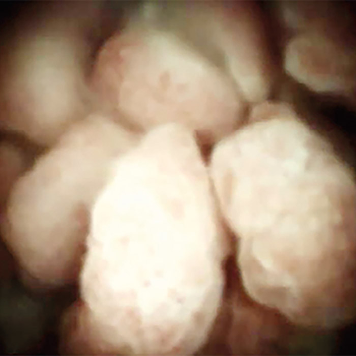
The SpyGlass DS System enables high resolution imaging and therapy during an endoscopic retrograde cholangiopancreatography (ERCP) procedure to target biopsies and fragment stones.1

​Previous research has demonstrated that visual impression of the stricture can also help guide patient management.2-4

A 2019 study examined the role of digital cholangiopancreatoscopy technology in mapping the extent of malignant involvement in the biliary or pancreatic ducts before surgical intervention. Through an analysis of 118 patients, it demonstrated that the surgical plan was altered in 34% of cases when using direct visualization with the SpyGlass™ DS System.5

85%

In a clinical study of 298 patients, clinical management was altered in 85% of patients undergoing diagnostic ERCP with Cholangioscopy.6

89%

Demonstrated 89% stone clearance after a single ERCP session may allow patients to avoid repeat endoscopic interventions or more invasive procedures to treat difficult biliary and pancreatic stones.7

buy

 In a study, it was suggested SpyGlass DS could help reduce number of repeat procedures, hence reducing the overall expenditure of hospitals.8

Clinical cases aren't predictive of device performance.

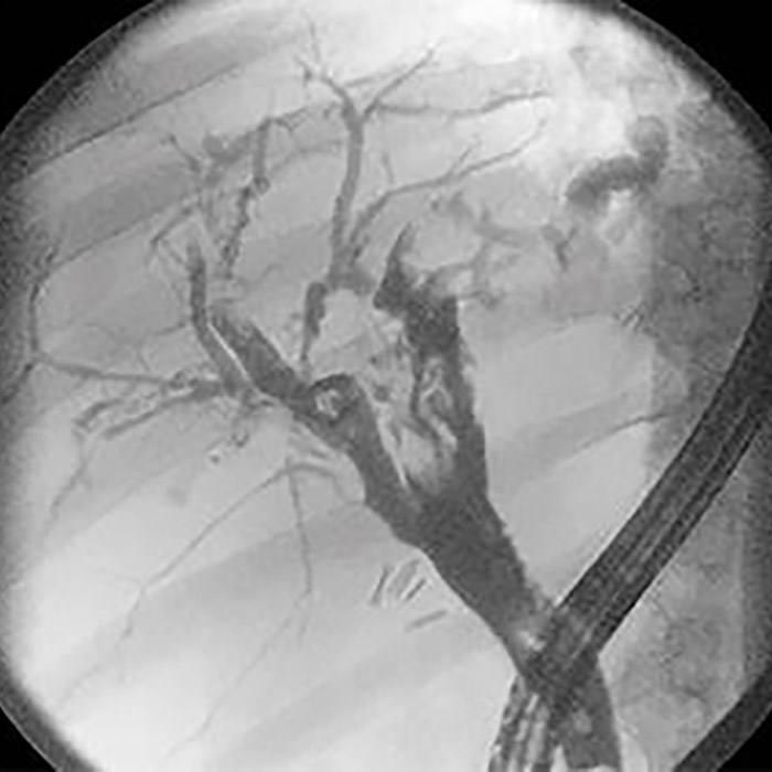
Diagnostic ERCP performed with:​

Fluoroscopic visualization​

Fluoroscopic visualization​

SpyGlass™ DS Direct Visualization System​

SpyGlass™ DS Direct Visualization System​

Find out how direct visualization can help you improve patient outcomes:

View product information for​ the SpyGlass™ DS Direct Vizualisation System:​

SpyGlass™  DS  Direct Vizualisation  System​

The SpyGlassTM Direct Vizualisation System enables direct visualization of the pancreatic and bile ducts. It is used to evaluate suspected benign and malignant conditions and to treat difficult stones and strictures​.

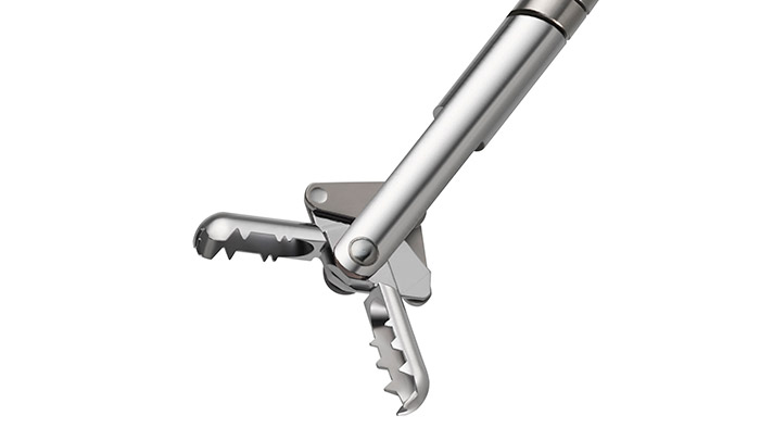
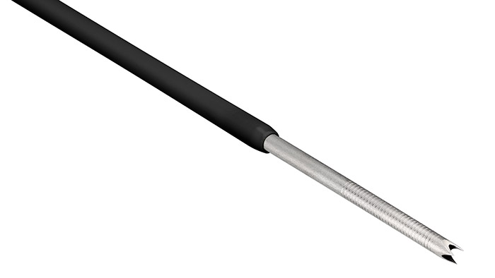
SpyGlass DS II clinical SpyBite

Driving innovation in HPB diagnosis​

At Boston Scientific, we understand the challenges you may face in achieving a diagnosis for your patients. We have developed a suite of solutions to help support them:​

Connect with us:


References

1. Draganov PV, Chauhan S, Wagh MS, Gupte AR, Lin T, Hou W, Forsmark CE. Diagnostic accuracy of conventional and cholangioscopy-guided sampling of indeterminate biliary lesions at the time of ERCP: a prospective, long-term follow-up study. Gastrointest Endos

2. Gerges C, Beyna T, Tang RSY, et al. Digital single-operator peroral cholangioscopy-guided biopsy sampling versus ERCP-guided brushing for indeterminate biliary strictures: a prospective, randomized, multicenter trial (with video). Gastrointest Endosc. 2020;91(5):1105–1113.​

​3. Laleman W, Verraes K, Van Steenbergen W, et al. Usefulness of the single-operator cholangioscopy system SpyGlass in biliary disease: a single-center prospective cohort study and aggregated review. Surg Endosc. 2017;31(5):2223–2232.​

4. Chen Y, Parsi MA, Binmoeller KF, et al. Single-operator cholangioscopy in patients requiring evaluation of bile duct disease or therapy of biliary stones (with videos). Gastrointest Endosc 2011;74(4):805–814.​

​5. Tyberg A, Raijman I, Siddiqui A, et al. Digital Pancreaticocholangioscopy for Mapping of Pancreaticobiliary Neoplasia: Can We Alter the Surgical Resection Margin? J Clin Gastroenterol. 2019;53(1):71–75.

6. Ramchandani et al. Single Operator Cholangioscopy for the evaluation and diagnosis of Indeterminate biliary strictures – Results from a Large Multinational Registry. DDW 2017.

7. Shah et al. Performance of a full disposable, digital, single-operator cholangiopancreatoiscope. Endoscopy 2017; 49; 651-658.

8. Deprez PH, Duran RG, Moreels T, Furneri G, Demma F, Verbeke L, et al. The economic impact of using single-operator cholangioscopy for the treatment of difficult bile duct stones and diagnosis of indeterminate bile duct strictures Authors. 2018; 109–118.

CAUTION: The law restricts these devices to sale by or on the order of a physician. Indications, contraindications, warnings, and instructions for use can be found in the product labelling supplied with each device or at www.IFU-BSCI.com. Products shown for INFORMATION purposes only and may not be approved or for sale in certain countries. This material not intended for use in France.​​All images owned by Boston Scientific.​