Boston Scientific accounts are for healthcare professionals only.


EPstar Fixed Electrophysiology Catheters
Configure or select a product to continue to order
- Overview
- Clinical data
- Technical specifications
- Ordering information
- Training
- Resources
Go further. Know more.™
Enhanced diagnostic precision for coronary sinus mapping and beyond
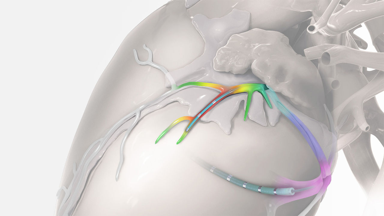
How it works
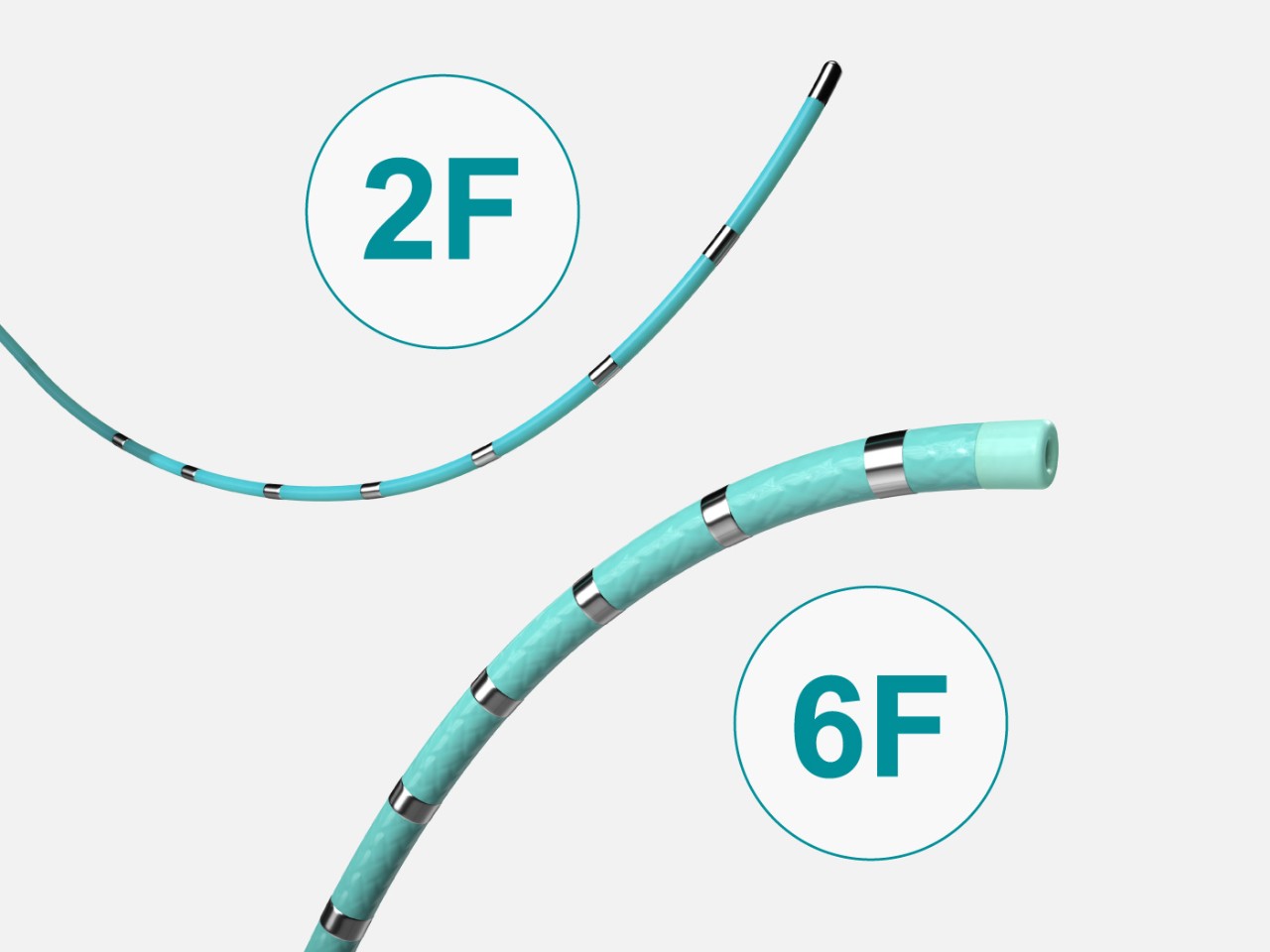
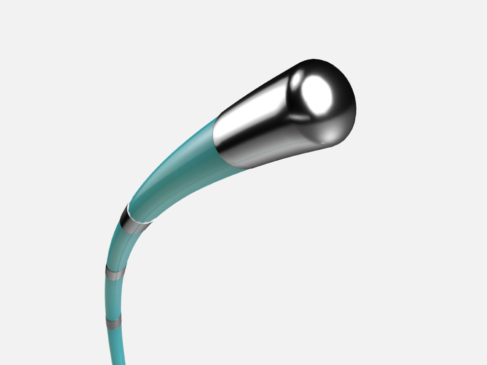
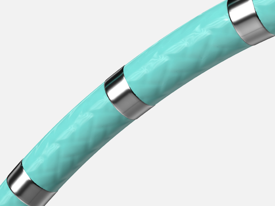
EPstar 2F Fixed Electrophysiology Catheter*
The 2F diagnostic microcatheter enables mapping and pacing in distal coronary sinus (CS) branches, which may be inaccessible to other catheters.
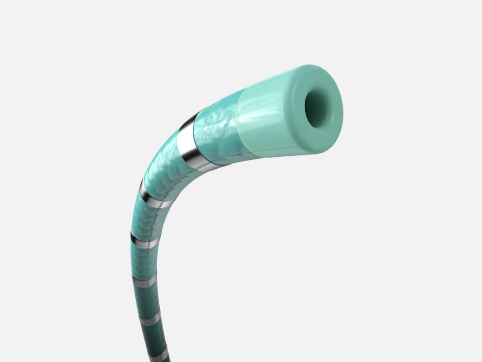
EPstar 6F Fixed Electrophysiology Catheter with Lumen†
The 6F diagnostic catheter is torquable with a large lumen, facilitating placement and telescoping of the 2F microcatheter in the coronary venous system.
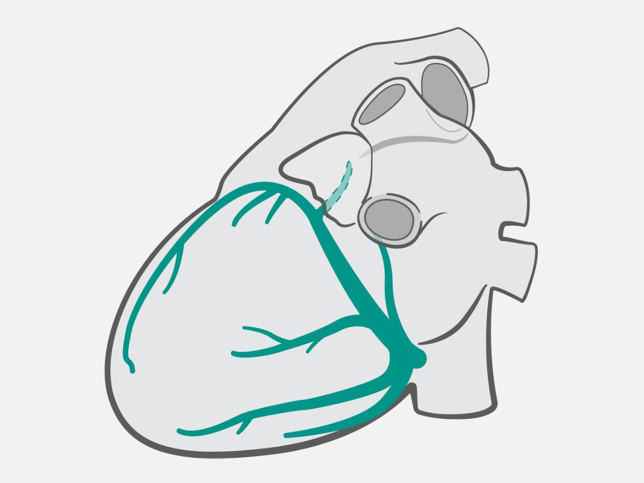
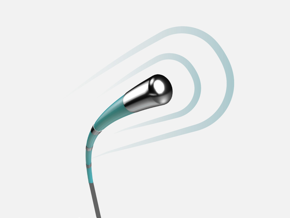
Using the EPstar 2F Microcatheter*
Enable mapping and pacing in distal coronary venous system branches with the octapolar* 2F microcatheter for deeper diagnostic precision
2F mapping has been used for:
- Idiopathic ventricular tachycardias1,2
- Complex atrial tachycardias3,4
- Left Wolff-Parkinson-White5
- Mapping and pacing in vein of Marshall3,6
Why choose EPstar Fixed Electrophysiology Catheters
Complex diagnostic mapping solutions
What’s included
EPstar Fixed Electrophysiology Catheters and Electrophysiology Cables are sold individually and as solution kits.
EPstar Coronary Venous Mapping Solution
- EPstar 2F Microcatheter*
- EPstar 6F Fixed Catheter with Lumen† (95 cm length)
- Connector cables
EPstar Bundle Kit (555)
- EPstar 2F Microcatheter*
- EPstar 6F Fixed Catheter with Lumen† (5-5-5 mm electrode spacing, 65 cm length)
EPstar Bundle Kit (282)
- EPstar 2F Microcatheter*
- EPstar 6F Fixed Catheter with Lumen† (2-8-2 mm electrode spacing, 65 cm length)


Related products

* EPstar Fixed Electrophysiology Catheter
† EPstar Fixed Electrophysiology Catheter with Lumen
References
- Komatsu, Y., et al. (2018). Idiopathic ventricular arrhythmias originating from the vicinity of the communicating vein of cardiac venous systems at the left ventricular summit. Circ Arrhythm Electrophysiol, 11(1). doi: 10.1161/CIRCEP.117.005386
- Ito, S., et al. (2005). Simultaneous mapping in the left sinus of valsalva and coronary venous system predicts successful catheter ablation from the left sinus of valsalva. Pacing Clin Electrophysiol, 28(s1). doi: 10.1111/j.1540-8159.2005.00081.x
- Kawamura, I., et al. (2019). Characteristics of Marshall bundle-related atrial tachycardias using an ultrahigh-resolution mapping system. J Interv Card Electrophysiol, 55(2), 161-169. doi: 10.1007/s10840-019-00544-9
- Yamamoto, T., et al. (2014). Marshall bundle reentry: A novel type of macroreentrant atrial tachycardia. Heart Rhythm, 11(7), 1229-1232. doi: 10.1016/j.hrthm.2014.03.051
- Davis, L., et al. (1995). Simultaneous mapping of the tricuspid and mitral valve annuli at electrophysiological study. Br Heart J, 73(4), 377-382. doi: 10.1136/hrt.73.4.377
- Fujisawa, T., et al. (2019). Importance of the vein of Marshall involvement in mitral isthmus ablation. Pacing Clin Electrophysiol, 42(6), 617-624. doi: 10.1111/pace.13640