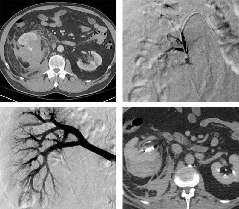
Renal pseudoneurysm embolization
Courtesy of Dr. Gary Siskin I Albany Medical Center
Presentation
60-year-old male patient who developed right flank pain and hematuria after falling. He became hypotensive and had a syncopal event. CT showed a large right retroperitoneal hematoma and a grade III renal laceration with evidence for active extravasation. He was transferred to our hospital where he became hypotensive with SBP in the 60s. He was transfused with 2u PRBC and referred for angiography and embolization.
Intervention used
Arterial access was gained via the right common femoral artery. A Sos-2 catheter was used to catheterize the right renal artery. Selective angiography demonstrated a pseudoaneurysm arising from the lower pole branch of the main renal artery. A Renegade™ HI-FLO™ Microcatheter was advanced into this vessel, and 0.2 mL of Obsidio Embolic was used for embolization. Follow-up angiography demonstrated successful occlusion of this vessel.
Outcome
He had no additional signs of ongoing bleeding with a stable hgb/hct after embolization. No additional transfusions were required. Follow-up CT demonstrated no further contrast extravasation and some retraction of the perirenal and retroperitoneal hematoma. He was discharged home 5 days after embolization.

Results from case studies are not necessarily predictive of results in other cases. Results in other cases may vary.
