Boston Scientific accounts are for healthcare professionals only.
IVUS 123 Essentials
IVUS elevates PCI outcomes.1 Clinical data consistently shows the benefits of IVUS to determine treatment strategy, guide stent placement, and assess procedural results. We developed IVUS 123 Essentials in partnership with physicians to simplify IVUS workflow and help improve outcomes for patients.
Data from the latest RENOVATE-COMPLEX-PCI Trial shows imaging-guided PCI led to a 37% reduction in event rates2 than angiography-guided PCI alone.3
IVUS 123 Essentials in Left Main PCI
See how Intravascular Ultrasound supports safe, effective, and durable Left Main PCI
What are the problems to be solved?
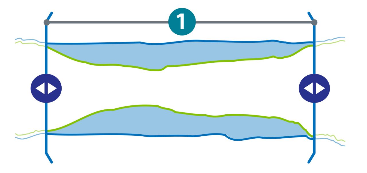
1. How long is the lesion to safely cover the plaque?
2. What is the plaque type and does it need modification prior to stenting?
3. What is the vessel size and what sized stent is required?
Pre-stent workflow
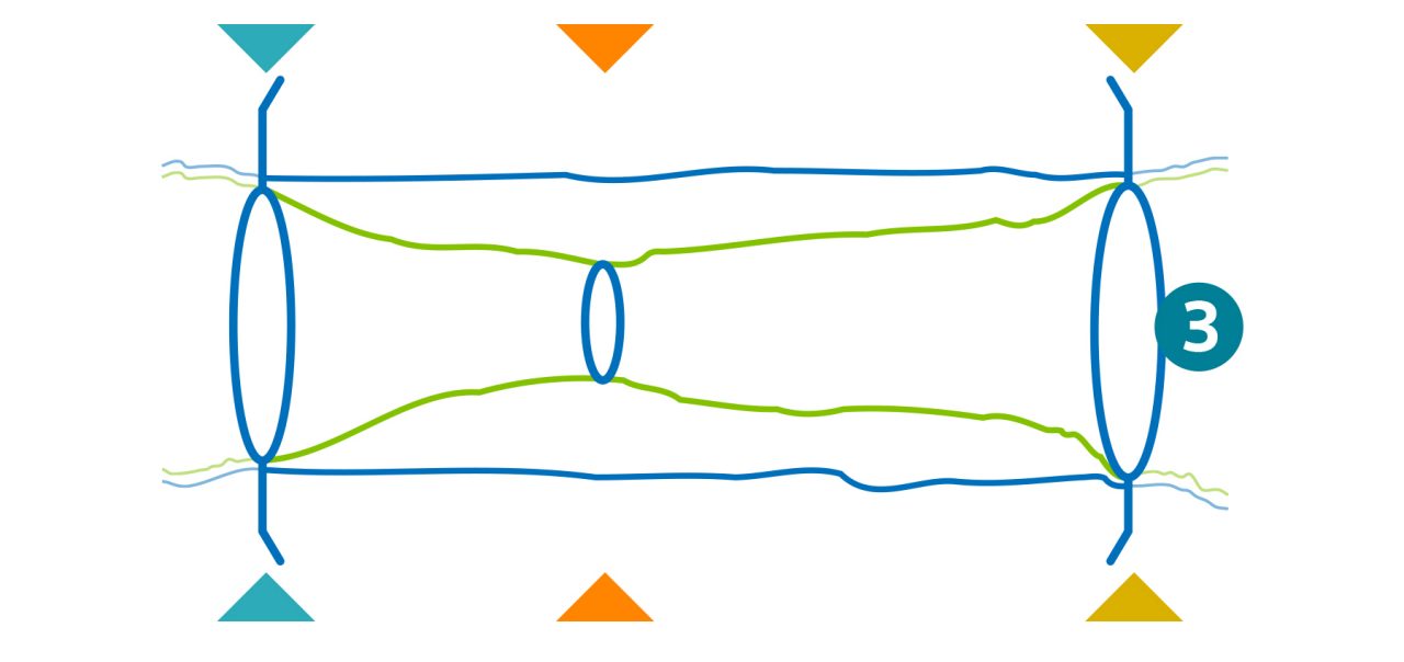
Take these 3 actions to determine treatment strategy.
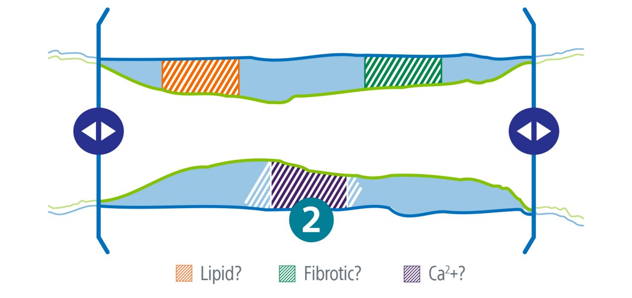
1. Establish lesion length and define landing zones.
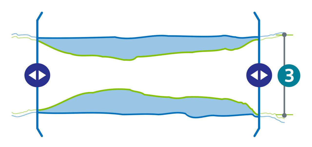
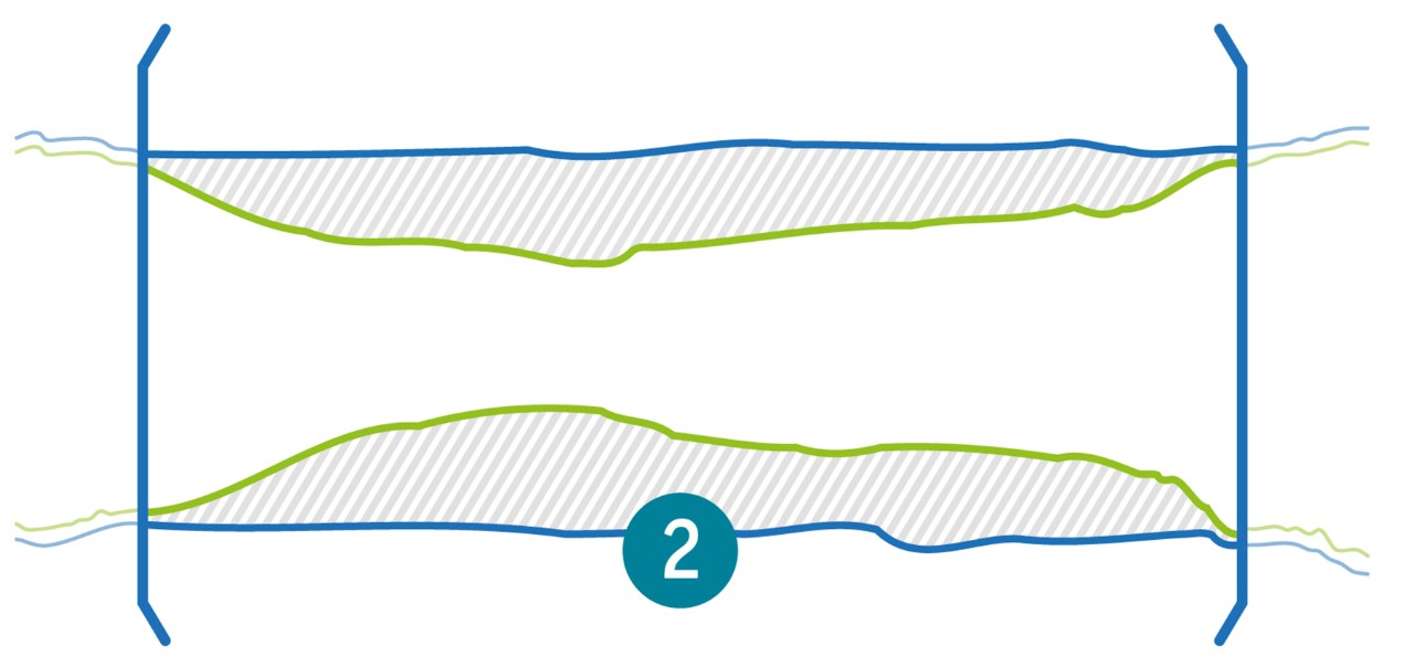
2. Assess plaque morphology.
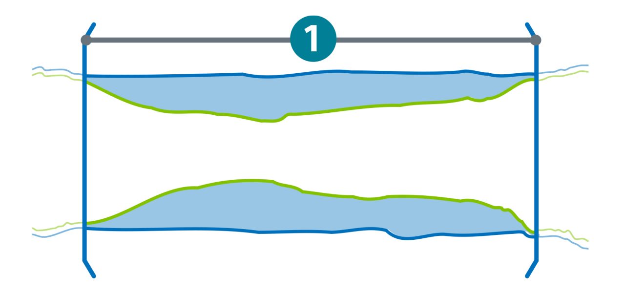
3. Measure the vessel size.
Post-stent workflow
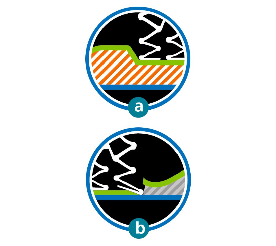
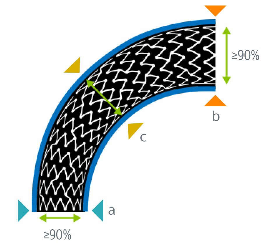
Ensure the best possible patient outcome by taking these final 3 actions:
1. Check for geographic miss (a) and edge dissection (b).
2. Check for malapposition.
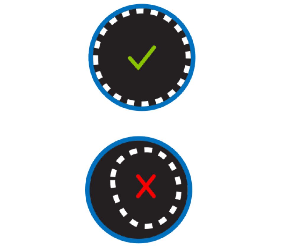
3. Check for optimal stent expansion.
Other IVUS resources
1. The SYNTAX II Trial studied the SYNERGY Bioabsorbable Polymer Stent in patients with either CTO or three vessel disease. The SYNTAX I trial studied the TAXUS Express2 drug eluting stent in patients with either Left Main or three vessel disease. Banning, Adrian, MBBS, MD. Clinical outcomes after state-of-the-art percutaneous coronary revascularization in patients with de novo three vessel disease: 5-year results of the SYNTAX II study. ESC Congress 2021.
2. Primary endpoint of cardiac death. target vessel-related Ml, or clinically driven target vessel revascularization
3. Lee, Joo Myung, et al. "lntravascular Imaging-Guided or Angiography-Guided Complex PCI." New England Journal of Medicine, vol. 388, no. 18, 5 Mar. 2023, pp. 1668-1679, https://doi.org/10.1056/nejmoa2216607
