SpyGlass™ DS
Direct Visualization System
Key Resources


Optimizing the Diagnosis and Treatment of Pancreaticobiliary Diseases:
Digital Cholangioscopy Using the SpyGlass™ DS System
Mouen Khashab, MD
Associate Professor of Medicine
Director of Therapeutic Endoscopy
Johns Hopkins Hospital
Baltimore, Maryland
Endoscopic retrograde cholangiopancreatography (ERCP) continues to be considered the standard method for the evaluation of pancreaticobiliary diseases.1 Peroral cholangiopancreatoscopy (POCPS) offers a valuable adjunct to ERCP for the visualization, diagnostic sampling, and treatment of biliary and pancreatic diseases.1-3 Recent technological advances have enhanced the visualization capabilities of POCPS using high-resolution digital technology.1,4
This monograph discusses the evolution of cholangiopancreatoscopy technologies and reviews the clinical data associated with the SpyGlass DS System, the most recent iteration of the technology.
Evolution of Direct Cholangioscopy
Although ERCP has been the standard technique for the nonsurgical diagnosis and management of pancreaticobiliary disease over the past several decades, this method relies on indirect visualization via fluoroscopic imaging of the biliary tree,5,6 and ERCP diagnosis of pathologic lesions via brush cytology and/or intraductal biopsy can have poor sensitivity.7 Consequently, many biliary lesions are indeterminate, which led to the development of a technique for direct visualization of the biliary tree for diagnosis and intervention—direct cholangioscopy.
Since the 1980s, several different strategies have been used for direct cholangiopancreatoscopy, including mother-daughter systems (insertion of a smaller cholangiopancreatoscope through a large-channel duodenoscope), and percutaneous cholangiopancreatoscopy through a mature T-tube tract.6,8 However, these strategies are limited by the need for 2 endoscopists, the fragility of the equipment, prolonged periods of time required for maturation of the T-tube tract, and poor visualization capabilities.8
Innovation in direct peroral cholangioscopy led to the development of the single-operator POCPS SpyGlass System SpyGlassTM Direct Visualization System (Boston Scientific Corporation), which was cleared for clinical use in the U.S. in 2005.9 The system consists of a catheter (SpyScope™) that is inserted through a standard working channel therapeutic duodenoscope.9 For visually directed biopsies, disposable biopsy forceps (SpyBite™) can be inserted into the SpyScope working channel.4
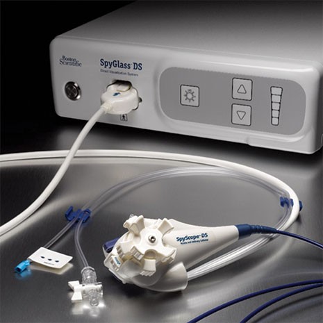
Recent technological advances have enhanced the visualization capabilities of the SpyGlass System using high-resolution digital technology, an improvement that also allows for a smaller image transmission cable, thereby improving maneuverability of the catheter tip. This new digital SpyGlass system—SpyGlass DS Direct Visualization System, made available for clinical use in 2015—has 2 dedicated irrigation channels and 4-way tip deflection.9,10 It includes a simplified setup using an integrated light source and camera (Figure 1). The device eliminates the need for reprocessing of the optical bundle as is required with the legacy SpyGlass.4 In addition, the digital optics along with improved suction and irrigation abilities enable substantially improved visualization.4
Potential diagnostic applications for the SpyGlass DS System include evaluation of indeterminate strictures; diagnosing indeterminate filling defects in the bile ducts observed during ERCP imaging; preoperative mapping of the precise location and extension of tumors of the pancreatobiliary tract; visual evaluation and biopsy of biliary stenosis; diagnosis of intraductal neoplasms; hemobilia; and detection and characterization of stones. Therapeutic applications include visually guided treatment of biliary stones that have otherwise failed extraction using conventional ERCP techniques, management of residual or impacted stones, removal of migrating biliary or pancreatic duct stents, and challenging cases of guidewire placement.2,3,11 In particular, the accuracy and operational efficiency of the SpyGlass™ DS System for the diagnosis and management of biliary and pancreatic disease is supported by extensive clinical data.
Biliary Stones
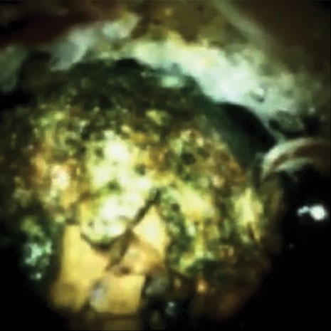
Complicated biliary stones include those that failed removal by mechanical lithotripsy, are impacted, or are wider than the diameter of the distal common bile duct.12,13 Such stones are more challenging to clear and may require more invasive removal techniques.12 Endoscopic laser lithotripsy (LL) and electrohydraulic lithotripsy (EHL) provide additional options for removal, but visualization of the target stone is important for accurate targeting, and this visualization can be achieved with cholangioscopy (Figure 2).12,14
Wong and colleagues published a prospective series evaluating the use of the SpyGlass DS System to guide visualization during the removal of complex biliary stones in 17 patients.13 Overall stone clearance was achieved in 16 of 17 cases (94%), and 63% of these were cleared in a single session.13 Cholangitis was the only adverse event reported, and occurred in 2 of the 17 cases (12%).13 The results of this study were notable given the patients’ median age of 76 years, the use in complicated cases, and the fast learning curve associated with the SpyGlass DS System.13
Brewer-Gutierrez and colleagues studied the utility of the SpyGlass DS system with EHL or LL in an international, multicenter study of 407 patients with obstructing biliary duct stones.15 Ducts were completely cleared in 97.3% of patients and were cleared in a single session in 77.4% of patients.15 Adverse events were reported in 15 patients (3.7%) which included cholangitis (6 cases), abdominal pain (5 cases), pancreatitis (1 case), bleeding (1 case), and bile duct perforation (1 case).15 Turowski and colleagues performed a retrospective, multicenter study of 107 SpyGlass DS procedures, and showed that biliary stones were visualized and completely removed in 91.1% of cases with a need for 3 procedures (range, 1-6) to achieve final stone clearance.16 Based on these data, the investigators concluded that interventions guided by the SpyGlass DS should be considered as the new standard approach for the diagnosis and therapy of large bile duct stones.16
Treatment results from the 10 studies that reflect stone removal treatments in 679 patients are summarized in the Table. Taken together, the published rate of stone removal was 95.6%.13,15,17-24 Of note, many of the study patients had large, complex, impacted stones that had been unsuccessfully treated with standard ERCP, and 63% to 88% of stones were removed in a single session, highlighting the potential advantage of SpyGlass™ DS System–guided lithotripsy to fragment and remove stones in a single procedure.13,20,23
A study by Mizrahi and colleagues compared EHL using the SpyGlass DS System with EHL using the legacy SpyGlass system for stone removal procedures (N=324).19 SpyGlass DS had a higher rate of stone clearance in the first procedure (83% vs 58%; P<0.01), shorter procedure time (49 vs 57 minutes; P=0.032), and reduced radiation doses (361 vs 620 mGy; P=0.02).19
Additional abstracts reported success with SpyGlass DS System–guided EHL in patients who previously failed ERCP for stone removal. In 5 abstracts and 466 combined cases, stones were successfully located in the common bile duct, common hepatic and cystic duct, and intrahepatic system. Rates of successful stone clearance using SpyGlass DS System–guided treatment ranged from 59% to 90%, and the overall clearance rate was 72% to 95%.23,26-29
Diagnosing and Mapping Biliary Strictures
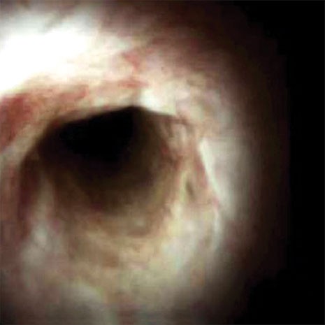
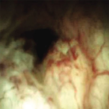
ERCP techniques alone are associated with suboptimal visualization of biliary and pancreatic duct stricture and suboptimal pathologic sensitivity and specificity of sampled lesions.30 POCPS can help overcome these limitations by enhancing direct visualization to obtain more accurate diagnostic sampling and by improving treatment of biliary and pancreatic strictures (Figures 3a and b).30
In a study of 117 cases with suspected malignancy, Turowski and colleagues reported that 99 cases could be stratified into benign (n=55) or malignant (n=44), indicating a sensitivity of 95.5% and a specificity of 94.5% for the diagnosis of malignancy.16 Based on these data, the investigators concluded that cholangioscopy with the SpyGlass DS System should be a new standard for the diagnosis of indefinite biliary lesions.16
In a prospective, multinational registry of POCS-guided biopsies using the legacy SpyGlass™ System and the SpyGlass DS System, investigators rated image quality as “excellent or good” in 76% of legacy cases compared with 96% of the SpyGlass DS System cases (P<0.001). The abstract indicated that, overall, image-based diagnostic accuracy with the SpyGlass DS System was 81%, and diagnostic accuracy from the SpyGlass DS System–guided SpyBite histopathology was 90%.31
Peroral cholangiopancreatoscopy can enhance direct visualization enabling the stratification of (a) benign and (b) malignant bile duct strictures.
In the largest published study to date using the SpyGlass DS System for diagnostic purposes, Shah and colleagues retrospectively collected data from 4 institutions in the United States.23 Eligible cases included 58 patients with indeterminate strictures.23 The SpyGlass DS System was associated with a sensitivity of 96.6% and a specificity of 93.3% for diagnosis by visual impression. Use of miniature biopsy forceps resulted in 86.2% sensitivity and 100% specificity.23 In addition to the high accuracy of visual and biopsy-based identification of pathology and detection of neoplasia, the investigators noted that the SpyGlass DS System improved optical resolution to clear biliary and pancreatic stones through a simplified setup.23
Ogura and colleagues used the SpyGlass DS System for diagnostic and therapeutic procedures in a prospective study of 55 patients with biliary disease and found that the diagnostic accuracy was 93%, with a sensitivity and specificity of 83% and 89%, respectively.21 The accuracy of forceps biopsy was 89%, and the sensitivity and specificity were 80% and 100%, respectively.21 The investigators concluded that diagnostic and therapeutic procedures using the new digital SpyGlass DS System were practicable and safe.21
Results from a retrospective study of 28 patients with biliary disorders showed the efficacy of the SpyGlass DS System for diagnosis in cases where ERCP had been unable to accurately detect abnormalities.18 The technical success rate for diagnostic procedures and the diagnostic accuracy were both 100%.18 The technical success rate for therapeutic procedures was 88%.18
In their comparative study of the SpyGlass DS System and the legacy SpyGlass device in 324 patients undergoing cholangioscopy for the diagnosis and management of pancreaticobiliary disease, Mizrahi and colleagues reported that the sensitivity for determination of malignant strictures was higher (78% vs 37%; P=0.004) and the procedure time was shorter (41 vs 51 minutes; P=0.03 minutes) for the SpyGlass™ DS System.19
In a study of 105 consecutive patients with suspected pancreatic or biliary disorders, Navaneethan and colleagues reported that in patients with indeterminate biliary strictures (n=44), it was possible to obtain clear views of the ductal lumen and mucosa in all cases using the SpyGlass DS System.20 Most procedures (74%) were conducted in an outpatient setting.20 Biopsies were adequate for histologic evaluation in 97.7% of patients, and the overall sensitivity and specificity for biopsy were 85% and 100%, respectively, using the SpyGlass DS System.20 The sensitivity and specificity for visual diagnosis of malignancy were 90% and 95.8%, respectively.20 The adverse event rate was 2.9%, with 2 cases of cholangitis and 1 case of post-procedural pancreatitis.20 The investigators noted that the SpyGlass DS System was easy to use and that its improved visualization and functionality could promote its use in the clinical setting.20
Tanaka and colleagues described their clinical experience using the SpyGlass DS System in diagnostic and therapeutic procedures in 26 patients with pancreaticobiliary disease, including those with indeterminate lesions of the biliary or pancreatic ducts.24 The overall technical success rate of visualizing the target lesions in cases with forceps biopsy was 100%, and the investigators concluded that digital cholangiopancreatoscopy can facilitate an accurate diagnosis and optimize treatment in patients with pancreaticobiliary diseases.24
Of note, authors of several clinical studies reported changing the planned treatment based on findings using the SpyGlass DS System. Such changes from previously planned treatment included identification of patients with unsuspected intrahepatic malignancy.23 In published abstracts, a study of 118 patients with pancreaticobiliary disorders used the SpyGlass DS System prior to planned procedures.32 In total, 39 patients (34%) had a change to their management plan based on the results, including avoidance of surgery in 28 (72%), change in type of surgery due to downstaging of cancer in 7 (18%), and upstaging of cancer in 4 patients (10%).32 This ability to accurately diagnose, target the proper treatment option, and potentially avoid unnecessary procedures through use of the SpyGlass DS System represents an important potential clinical advantage.
Furthermore, the SpyGlass™ DS System has clinical utility in pre-surgical planning. For example, in a case of a patient with intraductal papillary mucinous neoplasms (IPMN), the SpyGlass DS System localized the lesion in the main duct, permitting extended pancreatic tail resection instead of the previously planned total pancreatectomy.23 Ogawa and colleagues studied the use of the SpyGlass DS System to evaluate the longitudinal tumor extent in patients with cholangiocarcinoma, which is critical for any subsequent attempt at curative surgical treatment of extrahepatic cholangiocarcinoma.33 The investigators reported that the overall success rate for SpyGlass DS System–guided mapping biopsy was 88% and that the overall diagnostic accuracy for longitudinal tumor extent at the hepatic side, the duodenal side, and overall by SpyGlass DS System findings and mapping biopsy was 88% in all applications.33
| Clinical Parameters | Barakat et al, 2018 | Brewster Gutierrez et al, 2018 | Imanishi et al, 2017 | Mizrahi et al, 2018 | Navaneethan et al, 2016 | Ogura et al, 2017 | Ridtitid et al, 2017 | Shah et al, 2017 | Tanaka et al, 2016 | Wong et al, 2017 | Summary Outcomes |
| Therapeutic sample size | 39; 40 underwent procedure, but only 39 had stones or sludge found requiring treatment | 407 | 8 | 63 | 36 | 22 | 50 | 28 | 8 | 17 | 679 (678 underwent treatment; in 1 case, no stones were found) |
| Therapeutic success rate | 39 of 39 (100%) | 396 of 407 (97.3%) | 7 of 8 (87.5%) | 52 of 63 (83%) after first EHL | 35 of 36 (97.2%) | 20 of 22 (90.9%) | 45 of 50 (90%) | 28 of 28 (100%) | 7 of 8 (87.5%)b | 16 of 17 (94.1%) | 645 of 678 (95.1%) |
| Success rate of stone/sludge clearance | 39 of 39 (100%) | 396 of 407 (97.3%) | 4 of 4 (100%) | 52 of 63 (83%) after first EHL | 35 of 36 (97.2%) | 13 of 13 (100%) | 45 of 50 (90%) | 28 of 28 (100%) | 3 of 3 (100%) | 16 of 17 (94.1%) | 631 of 660 (95.6%) |
| Success of guidewire insertion/passage | NA | NA | 2 of 3 (66.7%) | NA | NA | 5 of 7 (71.4%) | NA | NA | 1 of 2 (50%) | NA | 8 of 12 (66.7%) |
| Success rate of migrating stent removal | NA | NA | 1 of 1 (100%) | NA | NA | 2 of 2 (100%) | NA | NA | 3 of 3 (100%) | NA | 6 of 6 (100%) |
| Adverse events | 3 of 40 (7.5%)
Pancreatitis (2)
Bleeding (1) | 15 of 407 (3.7%)
Cholangitis (6)
Abdominal pain (5)
Pancreatitis (1)
Bleeding (1)
Transient Bacteremia (1)
Bile duct perforation (1) | 0 of 8 (0%) | 2 of 126 (1.2%)a
Acute pancreatitis and abdominal pain (numbers not specified) | 3 of 105 (2.9%)a
Cholangitis (2)
Pancreatitis (1) | 1 of 22 (4.5%)
Cholangitis (1) | 5 of 50 (10%)
Pancreatitis (2)
Bleeding (2)
Cholangitis (1) | 3 of 108 (2.8%)a
Cholangitis (1)
Pancreatitis (1)
Abdominal Pain (1) | 2 of 26
(7.7%)a
Cholangitis (1)
Bleeding (1) | 2 of 17 (11.8%)
Cholangitis (2) | 36 of 909 (4.0%)a |
** Includes one additional patient not considered therapeutic in the publication because the initial goal was diagnostic. The patient then underwent a therapeutic intervention and therefore was considered in the therapeutic group for this analysis.
N=679
Based on references 13, 15, and 17-24.
Other Indications: Pancreatoscopy
In addition to its use in cholangioscopy, the SpyGlass DS System can facilitate successful pancreatoscopy. In a published abstract examining the role of the SpyGlass DS System in the evaluation of pancreatic IPMN, the SpyGlass DS System visualized villiform protrusions with a transparent appearance, villiform protrusions with a central vessel, nodules, luminal mucin, “fish-egg”¬–appearing side branches, and stones. Published abstracts showed that the system also determined more advanced disease in more than 50% of the patients with disease localized to the body of the pancreas, and extended the location of the tumor in 61% of patients when compared with their previous diagnosis.34 The SpyGlass DS System also has been shown to offer benefit in the evaluation of chronic calcific and recurrent pancreatitis, and cystic neoplasm.34,35
According to a recent study by Trindade and colleagues, the SpyGlass DS System identified findings that were not seen on cross-sectional imaging or endoscopic ultrasound in 42% of patients with main duct IPMN; subsequently, these findings dictated the type of surgery used in these cases.36 Furthermore, in patients with a diffusely dilated pancreatic duct (>10 mm), findings from the SpyGlass DS System dictated the type of surgery in 77% of these cases.36 Based on these data, the investigators concluded that digital pancreatoscopy with the SpyGlass DS System should be considered in the diagnostic algorithm of main duct IPMN in patients with a diffusely dilated pancreatic duct and without any focal lesions seen on cross-sectional imaging or endoscopic ultrasound.36
Safety and Reducing Fluoroscopy
Rates of adverse events in patients undergoing procedures assisted by the SpyGlass™ DS System were generally low and resolved with treatment.13,15,17-24 Adverse events included cholangitis, pancreatitis, abdominal pain, transient bacteremia, bile duct perforation, and bleeding (Table).13,15,17-24
Eliminating or reducing exposure to fluoroscopy-related radiation is an important goal for the safety of patients and health care providers. Furthermore, because fluoroscopy is generally not portable, its elimination means that otherwise unstable patients may be able to undergo diagnostic and therapeutic procedures without leaving the ICU.37 This strategy also could enable procedures in patients for whom exposure to radiation is undesirable (eg, pregnant women).38 In a study by Barakat and colleagues involving 40 patients, the SpyGlass DS System enabled fluoroscopy-free biliary cannulation in all patients (100%), with a median cannulation time of 64 seconds (range, 2 seconds to 15.4 minutes).17 In 2 cases (5%), 12 to 24 seconds of fluoroscopy was used just to confirm clearance of unexpectedly complex stones.17
Economic Advantages
Several groups of investigators have explored the cost-effectiveness of the SpyGlass DS System. Saumoy and colleagues constructed a decision analytic model to examine the cost and outcomes of strategies used to evaluate indeterminate strictures (eg, fluorescence in situ hybridization, probe-based confocal laser endomicroscopy, brush cytology, intraductal biopsy, and direct cholangioscopy).39 The abstract showed that, among these options, ERCP with cholangioscopy was the most cost-effective strategy.
In addition, using a model for management of difficult stones, Deprez and colleagues reported that the use of cholangioscopy was associated with a decrease in the number of procedures (–27% relative reduction) and costs (–€73,000; –11% relative reduction) when compared with ERCP.40 In a model of stricture diagnosis, the use of cholangioscopy was associated with a decrease in the number of procedures (–31% relative reduction) and costs (–€13,000; –5% relative reduction) when compared with ERCP.40
Castle and colleagues analyzed an open-label case series of 26 patients selected for digital single-operator cholangioscopy using the SpyGlass™ DS System during the first 18 months of implementation at a US hospital.41 The abstract indicated that the SpyGlass DS System resulted in a total net positive cost savings of $2,942 per patient over a 90-day follow-up after the procedure.41
Additional potential for cost-effectiveness has been demonstrated in clinical studies, but has not been studied in a formal model. In multiple studies, the use of the SpyGlass DS System was highly successful in guiding removal of complex stones that had previous treatment failures (Table).13,15,17-24 Given the high cost of medical procedures in patients who have known or suspected complexities, using the SpyGlass DS System at the time of initial treatment may increase the likelihood of successful treatment during the first session and may reduce the need for and subsequent costs of additional procedures. A reduced need for fluoroscopy also may decrease costs; however, formal studies have not been done to quantify the cost savings.
In other cases, surgical plans were altered or even cancelled due to the findings using the SpyGlass DS System.23,32,36 Additional studies that assess the impact of these avoided costs on disease management would further contribute to the cost-effectiveness calculation for the SpyGlass DS System. Other considerations for cost savings include the ease of setup and disposability of the device, which decreases cleaning and reprocessing time. These variables require further investigation.
Future Directions
Clinical data presented in this monograph hint at several future directions for the use of the SpyGlass DS System in the management of pancreaticobiliary disease. Evaluation of strictures may result in improved outcomes and reduced cost.16,40 Other investigators have concluded that the enhanced visualization capabilities of devices such as the SpyGlass DS System will initiate a shift to radiation-free interventions for biliary procedures that can be performed at the bedside.17
One group of researchers advocated establishing a standardized biopsy protocol when using the SpyGlass™ DS System. That group recommended taking at least 5 biopsies from one lesion to achieve a representative histology, increasing the shell of the biopsy forceps, or as demonstrated in a retrospective exploratory study, improving the diagnostic yield of the biopsies by using touch imprint cytology during SpyGlass DS System–guided biopsy.16,42
Accessories to broaden the utility of therapeutic cholangioscopy are currently under evaluation. Other investigators have advocated using chromoendoscopy in conjunction with cholangioscopy to evaluate mucosal lesions in the bile duct.43,44
Periprocedural antibiotics are likely useful for patients undergoing cholangioscopy. In the study by Turowski and colleagues, the rate of cholangitis was 12.8% without antibiotic prophylaxis and 1% after peri-interventional administration of antibiotics, indicating that the complication rate can be reduced considerably by using single-shot antibiotic treatment.16


