A challenging arteriovenous malformation of the external ear
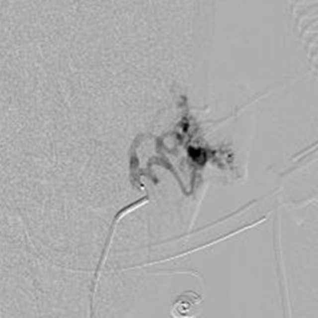
Baseline Central
A young girl with a serious Arteriovenous Malformation (AVM) of the left ear, principally filled by the auricolar artery, underwent angiographic treatment.
The angiographic evaluation showed two small vessels filling the nidus with a winding anatomy.
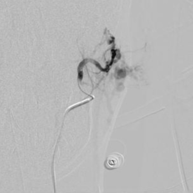
Embolization
After discarding any dangerous anastomoses which could have prevented the treatment, a Direxion™ Torquable Microcatheter and a Fathom™-16 guidewire were used to select distally one of those tiny branches.
Despite the high flow area we were able to cannulate the superior vessel and inject Glue.
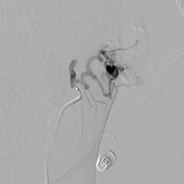
Result
In order to preserve the natural vascularization of the ear and prevent contrast and dose issues we decided to treat the inferior vessel in a second treatment.
Thanks to the handling of the microcatheter, obtained with the nitinol hypotube technology, we were able to easily engage and navigate in this tortuous anatomy.
Dr. Alfonso Marchianò – Chief of Interventional Radiology Department & Interventional Radiologist - IRCCS Istituto Nazionale dei Tumori" – Milano
Dr. Carlo Spreafico - Interventional Radiologist - "IRCCS Istituto Nazionale dei Tumori" – Milano